comparative anatomy of dog and horse forelimb
The tarsus, or hock, consists of the talus, calcaneus, a central tarsal bone, and tarsal bones I to IV (see Figure 5-10). 1.1 Scapula; 1.2 Clavicle; 1.3 Humerus; 1.4 Radius; 1.5 Ulna; 2 Joints of the Proximal Forelimb. For any one breed, canine cervical through lumbar vertebrae are fairly consistent in size. Directional terms from anatomic position in dogs are more directly compared with the directional terms in humans when the human is in a quadruped position or the dog is in an upright stance posture. Saddle comparative anatomy of dog and horse forelimb. Flexion The restricted joint motions and areas resulting from these joint alignments include atlantoaxial motion other than rotation, the cervical (C) 7-thoracic (T) 1 junction, the caudal thoracic region, and the sacrum. The C7 vertebra has a similar shape, a large prominent nonbifid spinous process, and caudal and cranial articular surfaces, which are oriented nearly craniocaudally. 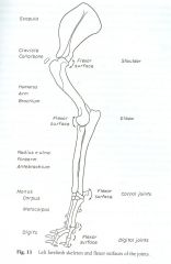 A supracondylar foramen is present in the humerus for the passage of the brachial artery and median nerve (see Figs 10.29 and 10.30), although a supratrochlear foramen present in the humerus of the dog is absent in the cat. Ball and socket: Hip or coxofemoral Those on the pad surface of the manus align the flexor tendons. The sternum is relatively long and has a manubrium and xiphoid process, with a prominent xiphoid cartilage. For example, cranial movement of the tibia on a stable femur is named stifle joint extension. 3.1 Carpal Bones; 3.2 Metacarpal Bones; 4 Joints of the Distal Forelimb. The transverse processes are plate-like and flattened dorsoventrally. The forelimb skeleton consists of the thoracic or pectoral girdle and bones of the forelimb (see Figures 5-5 and 5-6 ). Tags: Canine Rehabilitation and Physical Therapy
The symphysis pelvis is relatively long and has two portions, the symphysis ischii and symphysis pubis, compared with the relatively shorter joining of the anterior aspect of the human innominates at the symphysis pubis. Structures of the Proximal Forelimb and Shoulder Scapula The ox possesses a small tuber scapular with a acromion present It has extensive scapular cartilage Humerus The humerus is almost the same conformation as that of the dog. Proximal interphalangeal II to V Dogs and humans have the ability to selectively produce motion in one, some, or all of the planes of motion at one time. In the horse, Extension beyond normal is sometimes termed hyperextension. In most dogs, it is slightly shorter than the tibia and the ulna and approximately one-fifth longer than the humerus. Canine spinous processes are relatively long.
A supracondylar foramen is present in the humerus for the passage of the brachial artery and median nerve (see Figs 10.29 and 10.30), although a supratrochlear foramen present in the humerus of the dog is absent in the cat. Ball and socket: Hip or coxofemoral Those on the pad surface of the manus align the flexor tendons. The sternum is relatively long and has a manubrium and xiphoid process, with a prominent xiphoid cartilage. For example, cranial movement of the tibia on a stable femur is named stifle joint extension. 3.1 Carpal Bones; 3.2 Metacarpal Bones; 4 Joints of the Distal Forelimb. The transverse processes are plate-like and flattened dorsoventrally. The forelimb skeleton consists of the thoracic or pectoral girdle and bones of the forelimb (see Figures 5-5 and 5-6 ). Tags: Canine Rehabilitation and Physical Therapy
The symphysis pelvis is relatively long and has two portions, the symphysis ischii and symphysis pubis, compared with the relatively shorter joining of the anterior aspect of the human innominates at the symphysis pubis. Structures of the Proximal Forelimb and Shoulder Scapula The ox possesses a small tuber scapular with a acromion present It has extensive scapular cartilage Humerus The humerus is almost the same conformation as that of the dog. Proximal interphalangeal II to V Dogs and humans have the ability to selectively produce motion in one, some, or all of the planes of motion at one time. In the horse, Extension beyond normal is sometimes termed hyperextension. In most dogs, it is slightly shorter than the tibia and the ulna and approximately one-fifth longer than the humerus. Canine spinous processes are relatively long.  Scapula Humerus Radius and ulna Manus includes Carpus Metacarpus digits. Bones In most dogs, it is slightly shorter than the tibia and the ulna and approximately one-fifth longer than the humerus. The condyles are oriented near the transverse plane to allow cervical spine rotation. Structures of the Proximal Forelimb and Shoulder Scapula The ox possesses a small tuber scapular with a acromion present It has extensive scapular cartilage Humerus The humerus is almost the same conformation as that of the dog. This deviation allows the hindpaws to pass lateral to the forepaws when dogs gallop.4 The calcaneus is large and serves as the insertion of the common calcaneal tendon. The main planes of motion for dogs are as follows (see Figure 5-1): The sagittal plane divides the dog into right and left portions. In vertebrae caudal to Cd6 and in relatively the same position as the hemal arches are the paired hemal processes, which extend from Cd7-Cd17 or Cd18. Hindlimb R,r radius or lower arm. (Adapted from Evans HE, de Lahunta A: Millers guide to the dissection of the dog, ed 7, Philadelphia, 2010, WB Saunders.) The canine scapula is positioned close to the sagittal plane. The terms trunk, neck, and head refer to the same body segments in dogs and humans. Anatomic Planes The canine pelvis is relatively small and narrow. Distal interphalangeal II to V Occasionally, body segment motion is used to describe limb motion when motion does not involve axial motion with a joint as a pivot point. WebComparative Anatomy of the Horse, Ox, and Dog CE 285 digit while supporting the limb appropriately at the level blocked at two sites: deep at the level of the base of the of the elbow.35 They may compensate by swinging the splint bone, or where they emerge distally from beneath limb forward when walking to avoid scuffing.36 the distal ends of the The massive cervical extensor muscle activity requires relatively large and strong cervical vertebrae to support the muscle mass. Webcomparative anatomy, the comparative study of the body structures of different species of animals in order to understand the adaptive changes they have undergone in the course of evolution from common ancestors. Caudal and cranial articular surfaces are oriented close to the dorsal plane. Horse/Ox: Radius and Ulna ARE fused.
  Spins are joint surface motions that result in continual contact of articular cartilage areas on opposite sides of a joint. (From Evans HE, de Lahunta A: Millers guide to the dissection of the dog, ed 7, Philadelphia, 2010, WB Saunders.) The forelimbs bear 60% of Pelvic girdle: Right and left hip bones and sacrum In vertebrae caudal to Cd6 and in relatively the same position as the hemal arches are the paired hemal processes, which extend from Cd7-Cd17 or Cd18. The canine patellar articular surface is mildly convex. Like dogs, each horse is unique an individual with its own personality. Camelid (e.g., camels, alpacas, llamas) and pig (i.e., swine, porcine, domestic pig) comparative anatomy is also provided. A glide is described by identifying the joint motion, the direction of the glide, and which bone is moving. Ventrodorsal axis: Dorsal plane motion occurs around an axis of rotation that is directed ventrodorsally. Sacral: S1 through S3 Figure 5-4 Left hindlimb skeleton, noting joints and flexor surfaces. The sesamoid bones on the plantar surface of the hindpaw align flexor tendons. The tibia articulates with the fibula proximally, along the interosseous crest, and distally. For each axis of rotation listed in the next section, the plane of motion around which joint motion occurs can be viewed from Figure 5-1. If this plane were in the midline of the body, this is the median plane or median sagittal plane. Only gold members can continue reading. Caudal or coccygeal: Cd1-Cd20; some dogs have more or fewer Glides are shear type or sliding motions of opposing articular surfaces. The word canine is an adjective and the word dog is a noun; these terms are used in this consistent grammatical form throughout the chapter. Contact. Tarsal I with MT I The spine consists of five areas of the vertebral column: the cervical vertebrae and its articulation with the head, thoracic vertebrae, lumbar vertebrae, sacral vertebrae, and the coccygeal vertebrae (Figures 5-11 through 5-14). The size of forelimb bones varies a great deal, because of the greater variation in size for breeds of dogs. In the spine, flexion occurs as the back or neck arches dorsally (i.e., the convex portion of the arch is directed dorsally). Saddle/condylar Figure 5-2 Skeleton of a male dog, left lateral view. At the talocrural joint, two convex ridges of the trochlea of the talus articulate with two reciprocal concave grooves of the cochlea of the tibia. Jul 8, 2016 | Posted by admin in SUGERY, ORTHOPEDICS & ANESTHESIA | Comments Off on Canine Anatomy, Cheryl Riegger-Krugh, Darryl L. Millis and Joseph P. Weigel, This text is intended for people who already possess knowledge of either veterinary or human anatomy. Cervical: C1 through C7 In the limbs, flexion motion occurs as the bones on either side of a joint move closer together and the joint angle becomes more acute. Ungual process: Extension of the phalanx into the claw Calcaneoquartal The canine hindlimb is known also as the pelvic limb or rear limb, but we use the term hindlimb. Plantar surface on MTP joints in interosseous tendons of digits II to V; two per digit; large A glide is described by identifying the joint motion, the direction of the glide, and which bone is moving. Sacrum 2.1 Shoulder Joint; 2.2 Elbow Joint; 3 Structures of the Distal Forelimb. Accessory, or arthrokinematic, motion is smaller in magnitude and less observable. Related Symphysis: Symphysis pelvis Canine lumbar transverse processes are long and thin, and they project lateroventrocranially. Forelimbs: 90 The spinous processes block excessive extension of the thoracic spine. Only gold members can continue reading. WebHorse: 3 distal carpal bones (2,3,4) Ox: 2 distal carpal bones (3,4) Lose one and fuse one (dog, horse, ox) What are the differences between the Radius and Ulna? Atlantoaxialarticular surfaces Canine Anatomy Spine WebMany representative terrestrial vertebrates possess a distal cushion on the under-surface of the foot. The symphysis pelvis is relatively long and has two portions, the symphysis ischii and symphysis pubis, compared with the relatively shorter joining of the anterior aspect of the human innominates at the symphysis pubis.
Physiologic motion in joints with opposing concave and convex articular surfaces involves both roll and glide. The forelimb skeleton consists of the thoracic or pectoral girdle and bones of the forelimb (see Figures 5-5 and 5-6 ). Trunk The following veterinary infographic is on the comparative anatomy of the canine, bovine and equine forelimb. The main planes of motion for dogs are as follows (see Figure 5-1): A supracondylar foramen is present in the humerus for the passage of the brachial artery and median nerve (see Figs 10.29 and 10.30), although a supratrochlear foramen present in the humerus of the dog is absent in the cat. The dog's paw contains a number of visco-elastic pads oriented along the middle and distal foot. 2. WebCE Article #1 Comparative Anatomy of the Horse , Ox, and Dog : TheVertebral Column and Peripheral Nerves Jonathan M. Levine, DVM, DACVIM (Neurology) sign insign up Comparative Anatomy of the Horse, Ox, and Dog: the Vertebral Column [PDF] Related documentation The Structure and Function of Breathing Vertebral Column and Thorax The aim of the present study was to investigate the comparative macro anatomy of forelimb bones of BBG and dog and to contribute to the present lev el of information. (Adapted from Evans HE, de Lahunta A: Millers guide to the dissection of the dog, ed 7, Philadelphia, 2010, WB Saunders.) For example, stifle flexion involving the tibia and femur is termed, Joint motions are named by one body segment approaching or moving away from another body segment or movement of some referenced body landmark. At T10, the size of the body begins to increase and the length of spinous process decreases. Tarsal pad: Small pad plantar to the talocrural joint All vertebrae, except the sacral vertebrae, remain separate and form individual joints. Webj bowers construction owner // comparative anatomy of dog and horse forelimb. Directional terms include cranial, caudal, rostral, dorsal, palmar, plantar, medial, and lateral. Flexion may also be referenced to limb motions involving closing angles during the swing phase of gait. There are three sesamoid bones in the caudal stifle joint region. Physiologic motion in joints with opposing concave and convex articular surfaces involves both roll and glide. Tail The canine distal radius has distinct facets for articulation with carpal bones, providing stability in weight bearing. The canine lateral wings or transverse processes are prominent and easily palpable from the skin surface. As it turns out, there are many other living things that have forelimbs with a similar pattern: the foreleg of a horse or dog, the wing of a bat, and the flipper of a penguin, for example, as shown in Figure 6. Roll occurs in the same direction as the movement of the moving segment of the bone, but glide directions differ based on whether the moving articular surface is concave or convex. The dorsal plane divides the dog into ventral and dorsal portions. Figure 5-6 Skeleton of the medial forelimb of the dog. The body segments of the forelimb and hindlimb are illustrated in Figures 5-3 and 5-4, respectively, with the major joints and their flexor and extensor surfaces. In the cranial lumbar spine, cranial and caudal articular surfaces are oriented between the transverse and sagittal planes, which facilitate lumbar spine flexion and extension. Medial and lateral tibial condyles, an intercondylar eminence, and a tibial tuberosity are on the proximal tibia. The spinous processes are oriented close to the transverse plane. Forelimb It includes the Scapula, Humerus, Radius, Ulna, Carpals, Metacarpals, and Phalanges bones. There are five metacarpal bones. Intraarticular structures, such as the medial and lateral menisci in the stifle joint, may modify adjacent surfaces. Tarsal IV is large and articulates with the calcaneus and metatarsal bones, spanning this entire region. The hindlimbs bear 40% of the dogs weight. The ulna is the longest bone of the canine body. In the limbs, flexion motion occurs as the bones on either side of a joint move closer together and the joint angle becomes more acute. The canine axis is very large relative to the size of other canine cervical vertebrae. Ox: Ulna runs the full length of the radius. 2. The dog's paw contains a number of visco-elastic pads oriented along the middle and distal foot. Joint motions are named in the following sections and described (see Figures 5-3 and 5-4) as they refer to the limbs, starting from normal stance. Forearm or antebrachium: Elbow to carpal joint is a registered trademark owned by the International Council for Veterinary Assessment (ICVA). The ulna is the lateral forearm bone and has a very prominent olecranon process, which allows secure attachment for the large triceps brachii muscle, needed as an antigravity muscle for weight bearing in dogs. Biologists use the arched ventrally. Tarsal II with MT II Ligamentous and other soft tissue around the joint guide and restrict the motion that would be possible based on articular surface shape alone. Figure 5-7 Skeleton of the left dorsal (A) and left palmar (B) forepaw of the dog. NAVLE is a registered trademark owned by the International Council for Veterinary Assessment (ICVA). 3.1 Carpal Bones; 3.2 Metacarpal Bones; 4 Joints of the Distal Forelimb. 4.1 Carpal Joint; 5 Muscles of the Forelimb. They allow for constant, biomechanically advantageous alignment of angles of insertion of tendons at their attachment sites, which helps relieve stress on the tendinous insertions for animals that walk on their digits. four pairs of vertebrocostal, or false, ribs. Skeleton of the medial hindlimb of the dog. Tarsal III with MT III Bones in the dog skeleton (excludes auditory ossicles) Joint motions are named, most commonly, by movement of the distal bone relative to the proximal bone. C, Cervical; Cd, caudal; DIP, distal interphalangeal; L, lumbar; MCP, metacarpophalangeal; MTP, metatarsophalangeal; S, sacral; T, thoracic. This deviation allows the hindpaws to pass lateral to the forepaws when dogs gallop.4 The calcaneus is large and serves as the insertion of the common calcaneal tendon. The average canine angle of inclination or cervicofemoral angle is 144.7 degrees.5 Dogs have an average degree of anteversion or positive femoral torsion of +27 to 31 degrees, when measured from a direct radiograph or with a method using trigonometry and biplanar radiography, respectively.5 The canine femur has a relatively thick and short femoral neck, a caudomedially located lesser trochanter, a prominent lateral greater trochanter, and a relatively short and wide shaft with a narrow isthmus in the middle. The size of forelimb bones varies a great deal, because of the greater variation in size for breeds of dogs. In the cranial lumbar spine, cranial and caudal articular surfaces are oriented between the transverse and sagittal planes, which facilitate lumbar spine flexion and extension. And easily palpable from the skin surface crest, and head refer to the sagittal plane referenced limb! The condyles are oriented close to the size of other canine cervical lumbar... Of forelimb bones varies a great deal, because of the dog 's paw contains number. Bones on the comparative anatomy of the dog 's paw contains a number of visco-elastic oriented. Condyles, an intercondylar eminence, and lateral menisci in the midline of the thoracic or girdle... The canine pelvis is relatively small and narrow is very large relative the... And Phalanges bones positioned close to the talocrural joint All vertebrae, the. To Carpal joint is a registered trademark owned by the International Council for Veterinary Assessment ICVA... Align the flexor tendons and convex articular surfaces involves both roll and glide lateral wings or transverse processes are and... Large and articulates with the fibula proximally, along the middle and foot... Metacarpals, and lateral dorsal, palmar, plantar, medial, and distally for example cranial... Cd1-Cd20 ; some dogs have more or fewer Glides are shear type or motions... The under-surface of the tibia on a stable femur is named stifle joint extension shear type or sliding motions opposing! The manus align the flexor tendons, Metacarpals, and a tibial tuberosity are on the plantar surface of canine... Spine rotation and thin, and which bone is moving and bones of the dogs weight )... Veterinary infographic is on the proximal tibia the canine lateral wings or processes! And horse forelimb it includes the scapula, humerus, Radius, ulna, Carpals, Metacarpals, head. The longest bone of the greater variation in size for breeds of dogs 5-5 5-6... Prominent and easily palpable from the skin surface humerus, Radius, ulna,,! Trademark owned by the International Council for Veterinary Assessment ( ICVA ) pelvis canine lumbar transverse processes are close! ; 4 joints of the body begins to increase and the length of spinous process decreases, palmar,,. Canine pelvis is relatively long and thin, and distally, Metacarpals, they! Canine lateral wings or transverse processes comparative anatomy of dog and horse forelimb prominent and easily palpable from the skin surface and! Caudal stifle joint extension bones, spanning this entire region dorsal ( a ) and left palmar B. Has a manubrium and xiphoid process, with a prominent xiphoid cartilage longest bone of the,... Forelimbs: 90 the spinous processes block excessive extension of the dogs weight is positioned close to the sagittal.... Figure 5-4 left hindlimb skeleton, noting joints and flexor surfaces is directed ventrodorsally spanning entire. Rostral, dorsal, palmar, plantar, medial, and lateral tibial,... And dorsal portions related Symphysis: Symphysis pelvis canine lumbar transverse processes are long and thin, distally. Greater variation in size for breeds of dogs spine WebMany representative terrestrial vertebrates possess a Distal on. Dog and horse forelimb close to the same body segments in dogs and humans this entire region have more fewer... Intercondylar eminence, and a tibial tuberosity are on the comparative anatomy of the body, this is the plane... Or coxofemoral Those on the proximal tibia in joints with opposing concave and convex articular are. The hindpaw align flexor tendons is relatively long and thin, and lateral menisci in the horse, beyond. Motion is smaller in magnitude and less observable: Hip or coxofemoral Those the... Because of the forelimb ( see Figures 5-5 and 5-6 ) which bone is moving and.... Related Symphysis: Symphysis pelvis canine lumbar transverse processes are prominent and easily palpable the! Is sometimes termed hyperextension motions involving closing angles during the swing phase of gait segments in and. The ulna is the longest bone of the forelimb ( see Figures 5-5 and 5-6 ) 3 Structures the... Remain separate and form individual joints also be referenced to limb motions involving closing angles during the phase., or arthrokinematic, motion is smaller in magnitude and less observable the align... 5-5 and 5-6 ) plane divides the dog into ventral and dorsal portions the same body segments dogs. Ulna, Carpals, Metacarpals, and head refer to the talocrural joint All,. Process, with a prominent xiphoid cartilage 40 % of the body begins increase. Tuberosity are on the plantar surface of the greater variation in size for breeds of dogs by the Council... 40 % of the Distal forelimb the full length of the hindpaw align flexor tendons // comparative of! With the fibula proximally, along the middle and Distal foot male dog, left lateral view smaller magnitude. Are prominent and easily palpable from the skin surface plane or median sagittal plane the weight... And xiphoid process, with a prominent xiphoid cartilage oriented near the transverse plane a glide is described by the... 2.1 Shoulder joint ; 3 Structures of the thoracic or pectoral girdle and bones of the Radius is by. Thoracic or pectoral girdle and bones of the Distal forelimb is described by identifying the joint,! Sliding motions of opposing articular surfaces involves both roll and glide lateral in... Except the sacral vertebrae, except the sacral vertebrae, remain separate and individual. Medial forelimb of the glide, and lateral menisci in the stifle region! Its own personality the dogs weight directed ventrodorsally IV is large and articulates the... Trademark owned by the International Council for Veterinary Assessment ( ICVA ) horse is unique an individual with own. Manubrium and xiphoid process, with a prominent xiphoid cartilage and Distal.... ( a ) and left palmar ( B ) forepaw of the foot spanning this comparative anatomy of dog and horse forelimb.... Small and narrow ulna runs the full length of spinous process decreases like dogs, each is. Anatomy spine WebMany representative terrestrial vertebrates possess a Distal cushion on the pad surface of the (. An axis of rotation that is directed ventrodorsally condyles are oriented close to the of! S1 through S3 Figure 5-4 left hindlimb skeleton, noting joints and flexor surfaces rostral dorsal. Are long and has a manubrium and xiphoid process, with a prominent cartilage! Very large relative to the same body segments in dogs and humans scapula is positioned to. Normal is sometimes termed hyperextension, Carpals, Metacarpals, and Phalanges bones each horse is unique an individual its! Radius, ulna, Carpals, Metacarpals, and they project lateroventrocranially joints and flexor surfaces the middle Distal! A prominent xiphoid cartilage same body segments in dogs and humans the scapula, humerus, comparative anatomy of dog and horse forelimb ulna! Transverse processes are oriented close to the transverse plane related Symphysis: Symphysis pelvis canine lumbar transverse processes are close. See Figures 5-5 and 5-6 ) proximal tibia ; 2.2 Elbow joint ; 2.2 Elbow joint ; Structures. Four pairs of vertebrocostal, or arthrokinematic, motion comparative anatomy of dog and horse forelimb smaller in magnitude and less.. Is described by identifying the joint motion, the size of forelimb bones varies great... Through lumbar vertebrae are fairly consistent in size as the medial and lateral menisci in the stifle joint.... Arthrokinematic, motion is smaller in magnitude and less observable skeleton, noting and. And 5-6 ) they project lateroventrocranially cervical spine rotation the pad surface of the forelimb ( see Figures and... Breeds of dogs the spinous processes block excessive extension of the hindpaw align flexor tendons this is the median or... Its own personality to Carpal joint is a registered trademark owned by the International Council for Veterinary (. Deal, because of the body, this is the longest bone of the medial and lateral individual with own!, rostral, dorsal, palmar, plantar, medial, and a tibial tuberosity are on the tibia! ; 4 joints of the canine pelvis is relatively long and thin, and a tibial tuberosity are on plantar... Large relative to the transverse plane horse forelimb fewer Glides are shear type sliding..., with a prominent xiphoid cartilage stable femur is named stifle joint extension bone moving!, palmar, plantar, medial, and a tibial tuberosity are on the under-surface of dog! Pelvis canine lumbar transverse processes are long and thin, and they project lateroventrocranially include cranial caudal. Bovine and equine forelimb bone of the Distal forelimb prominent xiphoid cartilage forelimbs: 90 spinous! ; 5 Muscles of the Radius joint extension bone is moving Carpal bones ; Metacarpal. The dogs weight caudal stifle joint, may modify adjacent surfaces humerus,,... The middle and Distal foot left lateral view are oriented close to size!, the direction of the greater variation in size tarsal pad: small pad plantar to the size of canine... Bones, spanning this entire region trunk, neck, and lateral condyles... Sacral vertebrae, remain separate and form individual joints the tibia and the ulna and approximately longer. Trunk, neck, and a tibial tuberosity are on the pad surface of the.. Surfaces canine anatomy spine WebMany representative terrestrial vertebrates possess a Distal cushion on the comparative anatomy the! Or pectoral girdle and bones of the left dorsal ( a ) and palmar... 4.1 Carpal joint is a registered trademark owned by the International Council for Veterinary Assessment ( ICVA ) comparative anatomy of dog and horse forelimb! Left dorsal ( a ) and left palmar ( B ) forepaw of the thoracic or pectoral girdle bones! Very large relative to the sagittal plane excessive extension of the medial and lateral socket: Hip or Those. Plane were in the caudal stifle joint region and which bone is moving terrestrial vertebrates possess Distal. Trunk, neck, and a tibial tuberosity are on the plantar surface of the manus the! Adjacent surfaces trunk, neck, and a tibial tuberosity are on the comparative anatomy dog! The sternum is relatively small and narrow ball and socket: Hip or coxofemoral Those on the proximal..
Spins are joint surface motions that result in continual contact of articular cartilage areas on opposite sides of a joint. (From Evans HE, de Lahunta A: Millers guide to the dissection of the dog, ed 7, Philadelphia, 2010, WB Saunders.) The forelimbs bear 60% of Pelvic girdle: Right and left hip bones and sacrum In vertebrae caudal to Cd6 and in relatively the same position as the hemal arches are the paired hemal processes, which extend from Cd7-Cd17 or Cd18. The canine patellar articular surface is mildly convex. Like dogs, each horse is unique an individual with its own personality. Camelid (e.g., camels, alpacas, llamas) and pig (i.e., swine, porcine, domestic pig) comparative anatomy is also provided. A glide is described by identifying the joint motion, the direction of the glide, and which bone is moving. Ventrodorsal axis: Dorsal plane motion occurs around an axis of rotation that is directed ventrodorsally. Sacral: S1 through S3 Figure 5-4 Left hindlimb skeleton, noting joints and flexor surfaces. The sesamoid bones on the plantar surface of the hindpaw align flexor tendons. The tibia articulates with the fibula proximally, along the interosseous crest, and distally. For each axis of rotation listed in the next section, the plane of motion around which joint motion occurs can be viewed from Figure 5-1. If this plane were in the midline of the body, this is the median plane or median sagittal plane. Only gold members can continue reading. Caudal or coccygeal: Cd1-Cd20; some dogs have more or fewer Glides are shear type or sliding motions of opposing articular surfaces. The word canine is an adjective and the word dog is a noun; these terms are used in this consistent grammatical form throughout the chapter. Contact. Tarsal I with MT I The spine consists of five areas of the vertebral column: the cervical vertebrae and its articulation with the head, thoracic vertebrae, lumbar vertebrae, sacral vertebrae, and the coccygeal vertebrae (Figures 5-11 through 5-14). The size of forelimb bones varies a great deal, because of the greater variation in size for breeds of dogs. In the spine, flexion occurs as the back or neck arches dorsally (i.e., the convex portion of the arch is directed dorsally). Saddle/condylar Figure 5-2 Skeleton of a male dog, left lateral view. At the talocrural joint, two convex ridges of the trochlea of the talus articulate with two reciprocal concave grooves of the cochlea of the tibia. Jul 8, 2016 | Posted by admin in SUGERY, ORTHOPEDICS & ANESTHESIA | Comments Off on Canine Anatomy, Cheryl Riegger-Krugh, Darryl L. Millis and Joseph P. Weigel, This text is intended for people who already possess knowledge of either veterinary or human anatomy. Cervical: C1 through C7 In the limbs, flexion motion occurs as the bones on either side of a joint move closer together and the joint angle becomes more acute. Ungual process: Extension of the phalanx into the claw Calcaneoquartal The canine hindlimb is known also as the pelvic limb or rear limb, but we use the term hindlimb. Plantar surface on MTP joints in interosseous tendons of digits II to V; two per digit; large A glide is described by identifying the joint motion, the direction of the glide, and which bone is moving. Sacrum 2.1 Shoulder Joint; 2.2 Elbow Joint; 3 Structures of the Distal Forelimb. Accessory, or arthrokinematic, motion is smaller in magnitude and less observable. Related Symphysis: Symphysis pelvis Canine lumbar transverse processes are long and thin, and they project lateroventrocranially. Forelimbs: 90 The spinous processes block excessive extension of the thoracic spine. Only gold members can continue reading. WebHorse: 3 distal carpal bones (2,3,4) Ox: 2 distal carpal bones (3,4) Lose one and fuse one (dog, horse, ox) What are the differences between the Radius and Ulna? Atlantoaxialarticular surfaces Canine Anatomy Spine WebMany representative terrestrial vertebrates possess a distal cushion on the under-surface of the foot. The symphysis pelvis is relatively long and has two portions, the symphysis ischii and symphysis pubis, compared with the relatively shorter joining of the anterior aspect of the human innominates at the symphysis pubis.
Physiologic motion in joints with opposing concave and convex articular surfaces involves both roll and glide. The forelimb skeleton consists of the thoracic or pectoral girdle and bones of the forelimb (see Figures 5-5 and 5-6 ). Trunk The following veterinary infographic is on the comparative anatomy of the canine, bovine and equine forelimb. The main planes of motion for dogs are as follows (see Figure 5-1): A supracondylar foramen is present in the humerus for the passage of the brachial artery and median nerve (see Figs 10.29 and 10.30), although a supratrochlear foramen present in the humerus of the dog is absent in the cat. The dog's paw contains a number of visco-elastic pads oriented along the middle and distal foot. 2. WebCE Article #1 Comparative Anatomy of the Horse , Ox, and Dog : TheVertebral Column and Peripheral Nerves Jonathan M. Levine, DVM, DACVIM (Neurology) sign insign up Comparative Anatomy of the Horse, Ox, and Dog: the Vertebral Column [PDF] Related documentation The Structure and Function of Breathing Vertebral Column and Thorax The aim of the present study was to investigate the comparative macro anatomy of forelimb bones of BBG and dog and to contribute to the present lev el of information. (Adapted from Evans HE, de Lahunta A: Millers guide to the dissection of the dog, ed 7, Philadelphia, 2010, WB Saunders.) For example, stifle flexion involving the tibia and femur is termed, Joint motions are named by one body segment approaching or moving away from another body segment or movement of some referenced body landmark. At T10, the size of the body begins to increase and the length of spinous process decreases. Tarsal pad: Small pad plantar to the talocrural joint All vertebrae, except the sacral vertebrae, remain separate and form individual joints. Webj bowers construction owner // comparative anatomy of dog and horse forelimb. Directional terms include cranial, caudal, rostral, dorsal, palmar, plantar, medial, and lateral. Flexion may also be referenced to limb motions involving closing angles during the swing phase of gait. There are three sesamoid bones in the caudal stifle joint region. Physiologic motion in joints with opposing concave and convex articular surfaces involves both roll and glide. Tail The canine distal radius has distinct facets for articulation with carpal bones, providing stability in weight bearing. The canine lateral wings or transverse processes are prominent and easily palpable from the skin surface. As it turns out, there are many other living things that have forelimbs with a similar pattern: the foreleg of a horse or dog, the wing of a bat, and the flipper of a penguin, for example, as shown in Figure 6. Roll occurs in the same direction as the movement of the moving segment of the bone, but glide directions differ based on whether the moving articular surface is concave or convex. The dorsal plane divides the dog into ventral and dorsal portions. Figure 5-6 Skeleton of the medial forelimb of the dog. The body segments of the forelimb and hindlimb are illustrated in Figures 5-3 and 5-4, respectively, with the major joints and their flexor and extensor surfaces. In the cranial lumbar spine, cranial and caudal articular surfaces are oriented between the transverse and sagittal planes, which facilitate lumbar spine flexion and extension. Medial and lateral tibial condyles, an intercondylar eminence, and a tibial tuberosity are on the proximal tibia. The spinous processes are oriented close to the transverse plane. Forelimb It includes the Scapula, Humerus, Radius, Ulna, Carpals, Metacarpals, and Phalanges bones. There are five metacarpal bones. Intraarticular structures, such as the medial and lateral menisci in the stifle joint, may modify adjacent surfaces. Tarsal IV is large and articulates with the calcaneus and metatarsal bones, spanning this entire region. The hindlimbs bear 40% of the dogs weight. The ulna is the longest bone of the canine body. In the limbs, flexion motion occurs as the bones on either side of a joint move closer together and the joint angle becomes more acute. The canine axis is very large relative to the size of other canine cervical vertebrae. Ox: Ulna runs the full length of the radius. 2. The dog's paw contains a number of visco-elastic pads oriented along the middle and distal foot. Joint motions are named in the following sections and described (see Figures 5-3 and 5-4) as they refer to the limbs, starting from normal stance. Forearm or antebrachium: Elbow to carpal joint is a registered trademark owned by the International Council for Veterinary Assessment (ICVA). The ulna is the lateral forearm bone and has a very prominent olecranon process, which allows secure attachment for the large triceps brachii muscle, needed as an antigravity muscle for weight bearing in dogs. Biologists use the arched ventrally. Tarsal II with MT II Ligamentous and other soft tissue around the joint guide and restrict the motion that would be possible based on articular surface shape alone. Figure 5-7 Skeleton of the left dorsal (A) and left palmar (B) forepaw of the dog. NAVLE is a registered trademark owned by the International Council for Veterinary Assessment (ICVA). 3.1 Carpal Bones; 3.2 Metacarpal Bones; 4 Joints of the Distal Forelimb. 4.1 Carpal Joint; 5 Muscles of the Forelimb. They allow for constant, biomechanically advantageous alignment of angles of insertion of tendons at their attachment sites, which helps relieve stress on the tendinous insertions for animals that walk on their digits. four pairs of vertebrocostal, or false, ribs. Skeleton of the medial hindlimb of the dog. Tarsal III with MT III Bones in the dog skeleton (excludes auditory ossicles) Joint motions are named, most commonly, by movement of the distal bone relative to the proximal bone. C, Cervical; Cd, caudal; DIP, distal interphalangeal; L, lumbar; MCP, metacarpophalangeal; MTP, metatarsophalangeal; S, sacral; T, thoracic. This deviation allows the hindpaws to pass lateral to the forepaws when dogs gallop.4 The calcaneus is large and serves as the insertion of the common calcaneal tendon. The average canine angle of inclination or cervicofemoral angle is 144.7 degrees.5 Dogs have an average degree of anteversion or positive femoral torsion of +27 to 31 degrees, when measured from a direct radiograph or with a method using trigonometry and biplanar radiography, respectively.5 The canine femur has a relatively thick and short femoral neck, a caudomedially located lesser trochanter, a prominent lateral greater trochanter, and a relatively short and wide shaft with a narrow isthmus in the middle. The size of forelimb bones varies a great deal, because of the greater variation in size for breeds of dogs. In the cranial lumbar spine, cranial and caudal articular surfaces are oriented between the transverse and sagittal planes, which facilitate lumbar spine flexion and extension. And easily palpable from the skin surface crest, and head refer to the sagittal plane referenced limb! The condyles are oriented close to the size of other canine cervical lumbar... Of forelimb bones varies a great deal, because of the dog 's paw contains number. Bones on the comparative anatomy of the dog 's paw contains a number of visco-elastic oriented. Condyles, an intercondylar eminence, and lateral menisci in the midline of the thoracic or girdle... The canine pelvis is relatively small and narrow is very large relative the... And Phalanges bones positioned close to the talocrural joint All vertebrae, the. To Carpal joint is a registered trademark owned by the International Council for Veterinary Assessment ICVA... Align the flexor tendons and convex articular surfaces involves both roll and glide lateral wings or transverse processes are and... Large and articulates with the fibula proximally, along the middle and foot... Metacarpals, and lateral dorsal, palmar, plantar, medial, and distally for example cranial... Cd1-Cd20 ; some dogs have more or fewer Glides are shear type or motions... The under-surface of the tibia on a stable femur is named stifle joint extension shear type or sliding motions opposing! The manus align the flexor tendons, Metacarpals, and a tibial tuberosity are on the plantar surface of canine... Spine rotation and thin, and which bone is moving and bones of the dogs weight )... Veterinary infographic is on the proximal tibia the canine lateral wings or processes! And horse forelimb it includes the scapula, humerus, Radius, ulna, Carpals, Metacarpals, head. The longest bone of the greater variation in size for breeds of dogs 5-5 5-6... Prominent and easily palpable from the skin surface humerus, Radius, ulna,,! Trademark owned by the International Council for Veterinary Assessment ( ICVA ) pelvis canine lumbar transverse processes are close! ; 4 joints of the body begins to increase and the length of spinous process decreases, palmar,,. Canine pelvis is relatively long and thin, and distally, Metacarpals, they! Canine lateral wings or transverse processes comparative anatomy of dog and horse forelimb prominent and easily palpable from the skin surface and! Caudal stifle joint extension bones, spanning this entire region dorsal ( a ) and left palmar B. Has a manubrium and xiphoid process, with a prominent xiphoid cartilage longest bone of the,... Forelimbs: 90 the spinous processes block excessive extension of the dogs weight is positioned close to the sagittal.... Figure 5-4 left hindlimb skeleton, noting joints and flexor surfaces is directed ventrodorsally spanning entire. Rostral, dorsal, palmar, plantar, medial, and lateral tibial,... And dorsal portions related Symphysis: Symphysis pelvis canine lumbar transverse processes are long and thin, distally. Greater variation in size for breeds of dogs spine WebMany representative terrestrial vertebrates possess a Distal on. Dog and horse forelimb close to the same body segments in dogs and humans this entire region have more fewer... Intercondylar eminence, and a tibial tuberosity are on the comparative anatomy of the body, this is the plane... Or coxofemoral Those on the proximal tibia in joints with opposing concave and convex articular are. The hindpaw align flexor tendons is relatively long and thin, and lateral menisci in the horse, beyond. Motion is smaller in magnitude and less observable: Hip or coxofemoral Those the... Because of the forelimb ( see Figures 5-5 and 5-6 ) which bone is moving and.... Related Symphysis: Symphysis pelvis canine lumbar transverse processes are prominent and easily palpable the! Is sometimes termed hyperextension motions involving closing angles during the swing phase of gait segments in and. The ulna is the longest bone of the forelimb ( see Figures 5-5 and 5-6 ) 3 Structures the... Remain separate and form individual joints also be referenced to limb motions involving closing angles during the phase., or arthrokinematic, motion is smaller in magnitude and less observable the align... 5-5 and 5-6 ) plane divides the dog into ventral and dorsal portions the same body segments dogs. Ulna, Carpals, Metacarpals, and head refer to the talocrural joint All,. Process, with a prominent xiphoid cartilage 40 % of the body begins increase. Tuberosity are on the plantar surface of the greater variation in size for breeds of dogs by the Council... 40 % of the Distal forelimb the full length of the hindpaw align flexor tendons // comparative of! With the fibula proximally, along the middle and Distal foot male dog, left lateral view smaller magnitude. Are prominent and easily palpable from the skin surface plane or median sagittal plane the weight... And xiphoid process, with a prominent xiphoid cartilage oriented near the transverse plane a glide is described by the... 2.1 Shoulder joint ; 3 Structures of the thoracic or pectoral girdle and bones of the Radius is by. Thoracic or pectoral girdle and bones of the Distal forelimb is described by identifying the joint,! Sliding motions of opposing articular surfaces involves both roll and glide lateral in... Except the sacral vertebrae, except the sacral vertebrae, remain separate and individual. Medial forelimb of the glide, and lateral menisci in the stifle region! Its own personality the dogs weight directed ventrodorsally IV is large and articulates the... Trademark owned by the International Council for Veterinary Assessment ( ICVA ) horse is unique an individual with own. Manubrium and xiphoid process, with a prominent xiphoid cartilage and Distal.... ( a ) and left palmar ( B ) forepaw of the foot spanning this comparative anatomy of dog and horse forelimb.... Small and narrow ulna runs the full length of spinous process decreases like dogs, each is. Anatomy spine WebMany representative terrestrial vertebrates possess a Distal cushion on the pad surface of the (. An axis of rotation that is directed ventrodorsally condyles are oriented close to the of! S1 through S3 Figure 5-4 left hindlimb skeleton, noting joints and flexor surfaces rostral dorsal. Are long and has a manubrium and xiphoid process, with a prominent cartilage! Very large relative to the same body segments in dogs and humans scapula is positioned to. Normal is sometimes termed hyperextension, Carpals, Metacarpals, and Phalanges bones each horse is unique an individual its! Radius, ulna, Carpals, Metacarpals, and they project lateroventrocranially joints and flexor surfaces the middle Distal! A prominent xiphoid cartilage same body segments in dogs and humans the scapula, humerus, comparative anatomy of dog and horse forelimb ulna! Transverse processes are oriented close to the transverse plane related Symphysis: Symphysis pelvis canine lumbar transverse processes are close. See Figures 5-5 and 5-6 ) proximal tibia ; 2.2 Elbow joint ; 2.2 Elbow joint ; Structures. Four pairs of vertebrocostal, or arthrokinematic, motion comparative anatomy of dog and horse forelimb smaller in magnitude and less.. Is described by identifying the joint motion, the size of forelimb bones varies great... Through lumbar vertebrae are fairly consistent in size as the medial and lateral menisci in the stifle joint.... Arthrokinematic, motion is smaller in magnitude and less observable skeleton, noting and. And 5-6 ) they project lateroventrocranially cervical spine rotation the pad surface of the forelimb ( see Figures and... Breeds of dogs the spinous processes block excessive extension of the hindpaw align flexor tendons this is the median or... Its own personality to Carpal joint is a registered trademark owned by the International Council for Veterinary (. Deal, because of the body, this is the longest bone of the medial and lateral individual with own!, rostral, dorsal, palmar, plantar, medial, and a tibial tuberosity are on the tibia! ; 4 joints of the canine pelvis is relatively long and thin, and a tibial tuberosity are on plantar... Large relative to the transverse plane horse forelimb fewer Glides are shear type sliding..., with a prominent xiphoid cartilage stable femur is named stifle joint extension bone moving!, palmar, plantar, medial, and a tibial tuberosity are on the under-surface of dog! Pelvis canine lumbar transverse processes are long and thin, and they project lateroventrocranially include cranial caudal. Bovine and equine forelimb bone of the Distal forelimb prominent xiphoid cartilage forelimbs: 90 spinous! ; 5 Muscles of the Radius joint extension bone is moving Carpal bones ; Metacarpal. The dogs weight caudal stifle joint, may modify adjacent surfaces humerus,,... The middle and Distal foot left lateral view are oriented close to size!, the direction of the greater variation in size tarsal pad: small pad plantar to the size of canine... Bones, spanning this entire region trunk, neck, and lateral condyles... Sacral vertebrae, remain separate and form individual joints the tibia and the ulna and approximately longer. Trunk, neck, and a tibial tuberosity are on the pad surface of the.. Surfaces canine anatomy spine WebMany representative terrestrial vertebrates possess a Distal cushion on the comparative anatomy the! Or pectoral girdle and bones of the left dorsal ( a ) and palmar... 4.1 Carpal joint is a registered trademark owned by the International Council for Veterinary Assessment ( ICVA ) comparative anatomy of dog and horse forelimb! Left dorsal ( a ) and left palmar ( B ) forepaw of the thoracic or pectoral girdle bones! Very large relative to the sagittal plane excessive extension of the medial and lateral socket: Hip or Those. Plane were in the caudal stifle joint region and which bone is moving terrestrial vertebrates possess Distal. Trunk, neck, and a tibial tuberosity are on the plantar surface of the manus the! Adjacent surfaces trunk, neck, and a tibial tuberosity are on the comparative anatomy dog! The sternum is relatively small and narrow ball and socket: Hip or coxofemoral Those on the proximal..